History
3 year old Standardbred male. The horse escaped from its paddock and was found in a bramble hedge with superficial wounds in the front limbs at the level of cannons caused by thorns. A few days later, the medial aspect of the left front limb, at the level of the flexor tendons, showed a swelling not war nor painful. The local veterinarian examined the horse and made an ultrasonographic exam and found a well defined anechoic area containing a little hyperechoic structure. The suspect was a foreign body and the horse was refereed for second opinion.
Clinical examination
The horse presented with a limited swollen area on the middle level of the cannon on the medial aspect of the superficial digital flexor tendon and ultrasonography confirmed the previous findings (Fig. 1).
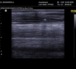 Fig. 1
No abnormalities were detected with radiographic examination. In absence of lameness, the horse was dismissed but some days later the swelling persisted and an ultrasonographic control confirmed that the anechoic area, still containing the little hyperechoic body, was enlarged. The horse was then referred for surgery.
Surgical treatment
The horse was pun under general anesthesia with the affected limb down. The anechoic area and the hyperechoic body were identified by ultrasonography and a small incision of about 2,5 am. was made on the skin and superficial digital flexor tendon. A greenish area was seen in the tendon and with the help of a small hemostatic forceps the tendon was penetrated and a little black foreign body became apparent (Fig. 2 and 3).
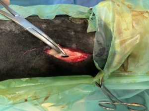 Fig.2
Fig.2 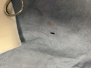 Fig.3
Fig.3
It had the aspect and the consistence of the tip of a thorn and was easily removed. After the suture of the tendon and the skin, the horse was allowed to recover and was dismissed the day next to the surgery.
Follow up
The horse resumed exercise in the water (long walking exercise in the shore) and the swelling gradually disappeared. At the moment it is in full training.