This branch is an important part of the clinic’s work.
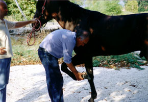
Horses admitted for lameness investigation are evaluated with the help of flexion tests and diagnostic anesthesia. In agreement with the owner and referring veterinarian, the diagnosis is completed with the help of diagnostic imaging, mostly radiology and ultrasonography.
The staff il also available for pre-purchase examinations
Diagnostic imaging
This is a continuously expanding sector, now calling for maximum reliability and specificity. Its benefits are reaped by all orthopaedic cases, clinically examined at Bagnarola or sent by colleagues for in-depth study. However, in many cases, diagnostic imaging also concern non-orthopaedic cases such as neurological disorders, subjects with thoracic or abdominal problems as well as problems affecting the uro-genital tract.
The diagnostic imaging department embraces: radiology, ultrasonography and nuclear scintigraphy.
Radiology
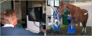
Radiography is still the most reliable investigating method, offering the greatest diagnostic detail. In spite of the advent of newer, more sophisticated methods, especially those focussed on investigating soft tissues in areas that cannot be reached with classic radiology, the radiographic examination represents a cornerstone of diagnostic imaging, and its present day evolution in the digital sphere enhances its versatility.
The Clinic has two radiological units, one of which is particularly high powered and thus able to examine areas that are usually inaccessible in the adult horse, such as the thorax, the thoracic-lumbar vertebral column, and the pelvis.
Ultrasonography
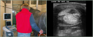
In the last twenty years, injuries to soft tissues - tendons and ligament in particular - have benefited from the advent and spread of diagnostic ultrasound. Today, ultrasonography provides an accurate diagnostic method, as well as monitoring of healing of orthopedic lesions.
Ultrasonography has proved to be a crucial tool also in diagnosing non-orthopaedic disorders, especially those affecting the abdomen and thorax.
Nuclear Scintigraphy
While radiology and ultrasonography give a kind of anatomical “photography” of lesions, scintigraphy offers a metabolic map of the skeleton, using a diphosphonate linked to a radioactive isotope that accumulates where there is a reparative process or bone remodeling.
The main indications for scintigraphy are: early diagnosis of “stress” fractures, especially in the racing horse; diagnosis of lameness positive to diagnostic blocks but without any radiological and ultrasonographic findings; diagnosis of “obscure” lameness of unidentified origin or located in areas not accessible to conventional methods.
