10 year old, Italian Showjumper mare.
History
The mare presented with history of recurrent colics, chronic weight loss, occasionally anorexia; recent weanling and non pregnant. Treated with omoprazole following a gastroscopic diagnosis of gastric ulcer. The colic had a sudden onset 9 hours before referral, with tachypnea, sweating and lateral recumbency.
Clinical examination
At admission the mare was moderately painful, T°C 37.9, HR 68 bpm, RR 10 rpm, and conjunctival mucous membranes markedly congested. On abdominal auscultation gut motility was absent. On rectal examination distended loops of small intestine were palpated. Gastric reflux was absent. Blood tests showed slightly increased PCV (44%), hypoprotidemia (6 gr/dL), WBC 6600/uL, blood pH 7.4 and BE 5.1. Two hours after admission clinical conditions worsened and the mare underwent exploratory laparotomy.
Treatment
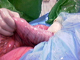
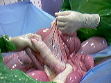
Exploratory laparotomy revealed moderately distended jejunum, whose lumen appears larger than normal with thickened but not edematous wall. The most distal part of the ileum had a more thickened wall probably secondary to hypertrophic muscular layer (Fig.1, 2).
 Fig.1
Fig.1  Fig.2
Fig.2
The intestinal lumen was nearly absent and attempts to empty the jejunum by massaging its contents in the caecum were inconclusive. The ileum was resected proximally to bypass the obstruction and a side-to-side jejunocaecal anastomosis was performed using GIA 80 stapling device. Intraopertively, a bioptic sample was taken off the wall of the ileum in its distal part for histopathology. The mare received antibiotic prophylaxis (Procain penicillin G 22.000 UI/kg IM and Gentamicin 6.6 mg/kg IV SID) for 5 days post op before being discharged.
Histopathology
Histopathology suggested primary hypetrophy of the ileum, but leiomyoma cannot be ruled out based on some cellular elements.
Follow up
Six months postoperatively the owner reported the mare in good health without relapse of colic.
