Direct inguinal hernia
A 5 year-old Thoroughbred was found in the morning with a swollen scrotum caused by a right sided inguinal hernia that the local veterinarian reduced manually per rectum.
In the evening the horse was still painless but with an enormously enlarged scrotum (Fig. 1).
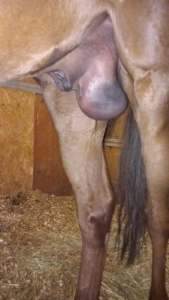 Fig. 1
At the time of admission the horse showed no signs of pain or distress. The right side of the scrotum was enlarged and on palpation it was difficult to find the right testicle whereas some intestinal loops could be appreciated under the skin.
The diagnosis was direct inguinal hernia and the surgical treatment was elected.
Following our routine anesthetic protocol, the horse was placed in dorsal recumbency and the inguinal area was surgically prepared. During this procedure the inguinal hernia was blindly reduced by gentle manual massage, facilitated by the dorsal recumbency.
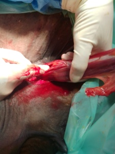
Using an inguinal surgical approach, the right testicle was isolated and externalized. The most proximal part of the vaginal tunic near the external inguinal ring showed a longitudinal tear with no evidence of intestinal loops (Fig. 2)
The right testicle was removed using a closed technique with ligature in correspondence of the most proximal border of the ruptured vaginal tunic. The external vaginal ring was closed using interrupted sutures, followed by continuous suture of the subcutaneous tissue and skin. The left testicle was removed following the routine technique.
The horse recovery was quick and smooth.
After 5 days the horse was discharged in good conditions with only a mild edema around the surgical wound.
The indirect inguinal hernia (with the intestine contained into the intact vaginal tunic) is the most common form in mature horses, while the direct inguinal hernia described in the present case is more frequent in foals.