History
Case 1 – 26 years old mare referred for esophageal obstruction after having been fed with carrots Case 2 – 7 years old pony female referred for esophageal obstruction occurred the evening before referral after eating pieces of apple.
Clinical examination and diagnostics
Both cases were referred after several vain attempts of managing the obstruction by siphoning with nasogastric tube. At admission, both horses appeared alert with some bilateral nasal discharge of saliva and no cough. In both cases further attempts of siphoning were performed – after administering metamizole and hyoscine as muscle relaxants – but it was not possible to advance the nasogastric tube over the proximal third of the esophagus: despite repeated flushing, the water flowed back clean and with no effect on the obstructing material. Contrast radiography was performed using liquid barium (Figs 1 and 2) to identify the nature and dimension of the obstructing material and to rule out abnormalities of the esophagus. In both cases the esophagus was normal.
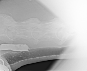 Fig.1
Fig.1 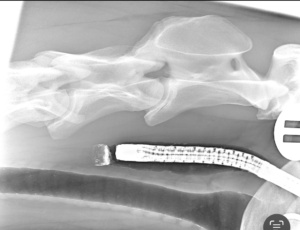 Fig.2
Fig.2
Using endoscopy, in the first case a piece of carrot was visible obstructing the whole lumen of the esophagus, while the pony appeared obstructed by material difficult to categorize.
In both cases – given the failure of the medical management – the surgical treatment was elected.
Surgery
Following our routine anesthetic protocol, both horses were placed in dorsal recumbency with the head extended, and an area extending from the larynx to the proximal third of the neck was surgically prepared. A nasogastric tube was inserted up to the obstruction to be used as a reference point. After incision of the skin and blunt separation of the paired sternothyroid, sternohyoid and omohyid muscles the esophagus was identified on the left side of the trachea and the nasogastric tube palpated. In both cases any manipulation and attempt to walk the obstructing material resulted impossible and so a longitudinal esofagotomy was performed. In the first case a piece of carrot was removed (Fig. 3), in the second case the obstructing body was a hard conglomerate of dehydrated hay and grass (Fig.4). In both cases the obstructing material was tightly fitting to the esophageal mucosa.
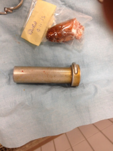 Fig. 3
 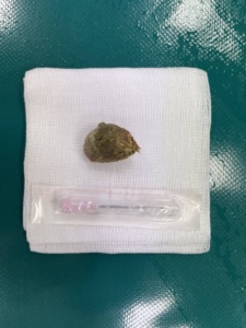 Fig.4
Fig.4
After removing the obstruction, the esophagus was sutured in two layers followed by the suture of the skin and application of a drain. Both horses recovered from anesthesia uneventfully. Perioperative antibiotics were used in both cases.
Follow up
Both horses were fasted for a day, then gradually returned to feeding and finally dismissed without complications after a week.