3 year old male Quarter Horse
History
The horse showed a 2/5 degree right front lameness after the second “go” in a reining competition. After radiographic examination, the local veterinarian diagnosed a fracture involving the whole base of the lateral sesamoid bone. The horse was referred to the clinic for fracture fixation.
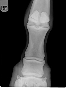 Fig. 1
Surgery
With the horse in lateral recumbency and the affected limb uppermost, a minimally invasive internal fixation was performed using a single 5,5 mm cortical screw in lag fashion under radiographic control. Initially a 4,5 screw was used but its purchase was considered poor, so it was replaced with a 5,5 screw. The limb was put in a cast and the horse recovered from anesthesia uneventfully.
The horse was dismissed after one week with the following program: 6 weeks box rest with cast, 4 weeks box rest with rigid bandage followed by 4 weeks box rest and then gradual resumption of exercise.
Follow up
The horse resumed regular work and shortly went back to its previous level of training. In a routine radiographic control, the screw was noticed to be partially displaced, protruding distally.
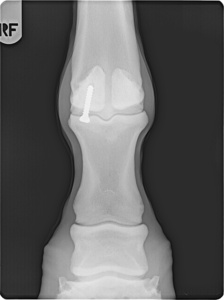 Fig.2
Fig.2
The horse was referred to the clinic where under general anesthesia the screw was easily removed. When back to competition, the horse resumed quickly its top level of performance and became the Italian champion in its discipline.