3 year old, Standardbred female.
History
Presented with history of mild recurrent colics during the last 2 months, successfully treated with FANS. The mare suffred a more dramatic episode of colic the day after a race, three weeks before admission. An antiulcer therapy with Omeprazole was started at the farm, without any improvement.
Clinical signs
Poor body condition. At admission, HR 28, RR 10, T°C 37.8, normal intestinal motility on all the abdominal quadrants, PCV 44%, TPP 7,6 gr/dL, WBC 5700/mL; no signs of abdominal discomfort. After 48 hours, the mare showed slight to moderate signs of abdominal pain after eating, without important alterations of physical and blood parameters. Pain resolved spontaneausly.
Treatment
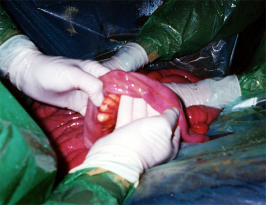
Exploratory laparotomy was performed.
Intraoperatively some fibrous blemish areas leading to stenosis of the intestinal lumen were detected on the ileal wall. A resection of 40 cm of ileum and a latero-lateral ileo-cecal anastomosis were performed. Samples of the resected segment were sent to the laboratory for histopathology.
Histopathology
Chronic inflammatory reaction on the muscular and submucosa layers of the ileal walls.
Follow up
During the early post operative period the mare didn’t show any sign of discomfort. Feeding was gradually started 24 hours post operatively. The mare was discharged 7 days post op, with instruction of keeping the mare on first quality hay and stall rest for 8 weeks.
Phone contact with the owner 30 days after discharge confirmed good general conditions of the mare which showed only a single episode of abdominal pain one time, which resolved with medical therapy.