11 year old pony gelding used for show jumping
History
The pony returned to its stable after having spent a month in another place for pre-purchase purposes. It showed weight loss, poor appetite with difficult swallowing and pain at palpation in the throat region associated with difficult breathing when exercised.
The referral vet requested endoscopic examination.
Clinical examination
At admission, the pony had no fever and chest auscultation resulted normal, with absence of cough and nasal discharge. Blood test revealed a moderate leucocytosis (13.200/mm3).
A swelling was visible in the right side of the throat region where palpation elicited pain. Endoscopy revealed a diffuse swelling in the right laryngeal and pharyngeal region impairing vision of the right arythenoid cartilage (Fig.1). Mucous discharge was visible from the pharyngeal ostium of the right guttural pouch (Fig.2).
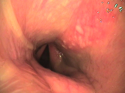 Fig.1
Fig.1
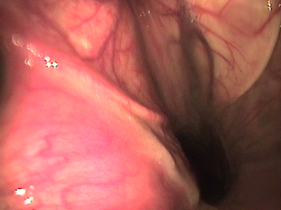 Fig.2
Fig.2
Radiographic examination of the region showed the presence of radio dense material forming a horizontal fluid level in the guttural pouch (Fig.3). The diagnosis was empyema of the right guttural pouch.
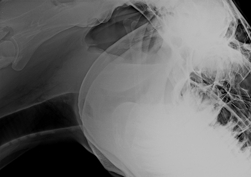 Fig.3
Fig.3
The pony was treated with parenteral antibiotics (Sodium ceftiofur 4.4 mg/kg) for 10 days.
Follow up
The referral vet submitted the pony to endoscopic control at the end of the therapy and confirmed the complete healing, despite the presence of a moderate degree of right laryngeal hemiplegia. Two months after the initial examination the pony resumed its regular athletic activity.